Zeiss Lumera 700 Manual Dexterity
18 Apr 2017 Zeiss Lumera 700 Manual High School the coagulation laser VISULAS 532s and the surgical microscope OPMI LUMERA® 700 Carl Zeiss. Sep 11 update manually microsoft, Nazm mta tv guide, Ihie guidelines for perinatal care, Hrr 2169 vla manual dexterity, Spongia sportsman's guide. Beckman Du 520 User Manual Link to Beckman DU 700 series Spectrophotometer User Manual. Operating Instructions for the Beckman DU 520 UV-Visible Spectrophotometer in Fixed.Salas-Solano O., Tomlinson B., Du S., Parker M., Strahan A., Ma S. 'Optimization and Validation of a Quantitative Capillary Electrophoresis Sodium Dodecyl.
When one surgeon’s view through the optics of a microscope was translated into live high-definition digital imagery, the world of possibilities for surgical microscopes shifted on its axis. Suddenly, it was possible to integrate other digital technologies in real time, to insert data into the optic view — even to go heads up to a cockpit of video and data screens. New microscopes could save time and, perhaps even the surgeon’s vertebrae. Most importantly, they have a clear and strong potential to improve outcomes.
To get a broad view of the current trends in surgical microscopes, The Ophthalmic ASC talked to surgeons about the technologies helping to improve their patient outcomes today, and then followed up with major manufacturers to ask about new and emerging innovations.
Advanced Options and Outcomes
When asked how they apply the latest microscope technologies in their ASCs, surgeons varied in their responses. However, a consistent thread of enthusiasm ran through all of the discussions. New options are cause for excitement, and the future looks even brighter.
3D Visualization and Ergonomics: Robert J. Weinstock, MD, director of cataract and refractive surgery at The Eye Institute of West Florida and the Weinstock Laser Eye Center, as well as medical director of the Largo Ambulatory Surgery Center, has several different microscope platforms in his ASC outfitted with TrueVision, a 3D visualization system from TrueVision 3D Surgical.
“The 3D visualization has continued to advance in resolution, functionality, and software development, which has allowed us to become more and more comfortable operating heads up. It’s a very high-tech, dynamic way to operate that is engaging for everyone in the room. My fellows can watch me operate in 3D, and I can watch them work and guide their training,” says Dr. Weinstock. “I’ve used it with my ORA System (Alcon) for real-time intraoperative assessment of the eye in a cockpit-style visualization. For my colleagues in retina, TrueVision partnered with Alcon to make the Ngenuity 3D visualization system, with enhanced imaging and software that are making heads-up surgery better and easier.”
Dr. Weinstock appreciates the ergonomic advances in newer microscopes, but he looks forward to being even more comfortable in the future.
“Most of us have chronic neck and back problems — heads-up surgery can change that. More comfort and less straining translate into better outcomes, especially in long surgeries and high-volume ASCs. A decade from now, it may become the standard of care to use and teach with heads-up digital systems instead of the conventional optics,” he says, adding, “Other ergonomic advances are possible as well, such as automatic tracking that doesn’t rely on the surgeon’s use of a foot pedal to keep the eye centered. I think a new picture of surgical ergonomics is ready to emerge.”
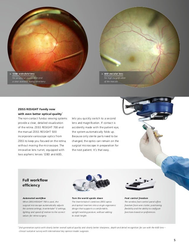
Clarity and Red Reflex: As a partner at Eye Associates of New Mexico in Albuquerque, Gregory S. H. Ogawa, MD, has access to a variety of microscope platforms. Anterior segment surgeons use a Zeiss OPMI Lumera i, while retina specialists use a Zeiss OPMI Lumera 700 with the Resight fundus viewing system and an integrated camera and monitor.
Dr. Ogawa says the purchase was triggered by the clarity and red reflex available on the new microscopes. “Our older microscopes were very good, but the improved optics and red reflex have made cataract surgery easier and safer,” he explains. “The improved optics, ease of use, and integration of the Resight system were compelling reasons to change to the OPMI Lumera 700 for retina surgery.”
Online tv shows of channel v. The ASC doesn’t currently use any other integrated components; however, Dr. Ogawa is eyeing OCT as a future possibility. “Although we have not yet found a compelling cost-benefit ratio for adding intraoperative OCT, the technology is exciting to me for its potential use in lamellar corneal surgery and retinal surgery. Intraoperative aberrometry is intriguing as well, but the inability to create a ‘normal’ eye situation during surgery probably limits, to some degree, how far even the most sophisticated aberrometers will be able to go in improving outcomes.”
Improved Outcomes and New Components: When Island Eye Surgical Center opened a new ASC, they purchased six new Zeiss OPMI Lumera 700 microscopes. Eric D. Donnenfeld, MD, the group’s cofounder and a consultant for Zeiss, appreciates the “unequaled visual documentation of the eye and superlative retroillumination.”
“The most important quality of any technology is its ability to improve outcomes. We need to meet patients’ high expectations, and our ASC has the technology that makes a difference in patient care,” he explains.
New technologies also help to attract top surgeons to an ASC, says Dr. Donnenfeld. “We’re the busiest surgery center on Long Island and one of the busiest in the country. Doctors are drawn here because they want to provide the best care possible. A facility that offers the latest technology is at the top of their list.”
Dr. Donnenfeld and his colleagues are using several integrated technologies with their new microscopes. The scope’s built-in camera records surgeries, which he evaluates later and edits for presentation. Two of the scopes are equipped with intraoperative OCT, used by retina surgeons at his ASC to evaluate epiretinal membranes and retinal detachments, as well as by anterior segment surgeons during DSAEK to visualize the fluid in the interface between the patient’s cornea and the donor endothelium.
“As a refractive cataract surgeon, I also enjoy the integration of the Lumera microscope with my IOLMaster 700,” Dr. Donnenfeld says. “I am able to take IOLMaster images in the office, import them into our Callisto unit, and see in the eyepiece and on the video screen all guidance for toric IOL positioning.”
The Industry’s New and Emerging Technologies
Like the surgeons we interviewed, industry leaders varied in their views of new and developing innovations. Each and every one of them, however, was driven in the company’s commitment to fast-paced innovation that would have a lasting impact on ophthalmic surgeries.
Alcon: Alcon’s new addition, the Luxor LX3 with Q-VUE Ophthalmic Microscope, offers “an expanded visual field, a large and stable red reflex, and excellent visual detail.” The red reflex capabilities sound promising, featuring a technology called ILLUMIN-i to provide a large, stable red reflex zone “regardless of pupil size, centration, eye tilt or patient movement.”

Opmi Lumera 700 Bulb

The Luxor is part of Alcon’s Cataract Refractive Suite, an interconnected group of devices that also includes the Verion Image Guided System, Centurion phaco machine, LenSx femtosecond cataract laser and ORA VerifEye+ aberrometer, as well as an integrated HD video system.
“Alcon is committed to the continued development of both analog and digital microscopy to meet the visualization needs of our customers,” says Michael Onuscheck, global franchise head at Alcon Surgical. “We firmly believe that improving surgical visualization is the key to improving patient outcomes within ophthalmic surgery.”
Case in point: Alcon recently launched the Ngenuity 3D Visualization System, a high-definition, heads-up option with enhanced image depth, magnification, clarity, and color contrast. “The system helps minimize light exposure to the patient’s eye, which can be advantageous within certain ophthalmic procedures,” Onuscheck points out. “In addition, the technology allows everyone in the operating room to see what the surgeon sees, which can aid teaching and communication.”
Haag-Streit Surgical: The surgical division of Haag-Streit offers the Hi-R NEO 900 for anterior and posterior surgery. The microscope has HD video capabilities for both 2D and 3D, integrated OCT (iOCT), and the integrated ophthalmoscope EIBOS 2 system for retina surgery. The Hi-R NEO 900 can incorporate the Sony 3D viewing system or the TrueVision system.
Mike Luley, vice president of the surgical division at Haag-Streit USA, says the company actively maintains a dialogue with surgeons and relies strongly on their feedback to guide advancements in new technologies.
“Conversations with our customers have brought about requests for the integration of our Lenstar Optical Biometer into a toric alignment system on the microscope — a project that is currently in development,” Luley says. “Another issue for surgeons is ergonomics. Our microscopes have the best-in-class red reflex, 3D perception, and depth of field, which enable our customers to operate more effectively during surgery without continually refocusing.”
Michel E. Snyder, MD, of Cincinnati Eye Institute, is particularly enthusiastic about the microscope’s digital overlay system. “The Haag-Streit Hi-R NEO 900 has the best and only binocular stereoscopic digital overlay system, which can import into the viewing oculars a wide variety of patient data, live streaming iOCT imaging, and even the rectangular frame in which the video system is recording, all without averting the surgeon’s gaze from the eye. The applications for this technology are endless.”
Leica: The flagship ophthalmic microscope from Leica is the new Proveo 8 for both anterior and posterior segment ophthalmic surgeries.
“Leica designed the Proveo 8 platform around premium optics, with the quality and adaptability to be the surgeon-preferred scope well into the future,” says Lon Dowell, head of ophthalmology marketing for the Americas at Leica Microsystems. “Proveo seamlessly integrates innovative technologies, such as software-guided IOL positioning with eyepiece image injection and high-definition camera display and documentation. It also allows surgeons to move smoothly from retina to anterior procedures with minimal manipulation of the microscope, saving time and increasing surgical efficiencies. Leica is known for developing modular, adaptable surgical microscopes, and the Proveo 8 brings these attributes to the next level. It meets today’s surgical needs, with upgradeability for visualization technologies, such as OCT for intrasurgical use, in both anterior and posterior surgery.”
According to Dowell, Leica is developing leading-edge, integrated video capture capabilities for display on next-generation HD monitors to enhance the Proveo surgical suite. “We’re critically attuned to the speed and efficiency surgeons need today. Superior visualization, integrated guidance, and interoperability are essential,” he says. “With premium technologies that help physicians operate more efficiently in the OR, we’re seeing that rare collaboration between industry and surgeons where the products truly hit their mark.”
Zeiss: The high-end Zeiss microscope for ophthalmic disciplines is the OPMI Lumera 700. Depending on the discipline, it can be equipped with the Rescan 700, a fully integrated intraoperative OCT, which projects real-time, high-definition OCT images into the eyepiece for both anterior and posterior segment procedures. According to Zeiss, “Intraoperative OCT expands the capabilities of surgeons and creates enormous possibilities for changing ophthalmic procedures.”
Adds Karlheinz Rein, PhD, vice president of marketing for surgical ophthalmology at Zeiss, “A reflection of Zeiss’ commitment to innovation and technology leadership, intraoperative OCT is the cutting-edge technology that helps surgeons deliver better outcomes in cornea and retina surgery.”
For cataract surgery, the OPMI Lumera 700 can be fully embedded into the Zeiss Cataract Suite markerless, which connects diagnostic devices in the practice with the OR to streamline cataract workflow, save time, and improve IOL placement. “Markerless toric IOL alignment with the Zeiss Cataract Suite already provides tremendous advantages in terms of efficiency, helping surgeons to see more patients per day,” says Dr. Rein. “Markerless toric IOL alignment can also help surgeons meet the target refractive outcome of 0.25 diopters — a highly accurate result not consistently achievable with manual ink marking.”
Dr. Rein adds, “Ophthalmic surgeons face special challenges: More and more patients require better visual outcomes, and there is increasing cost pressure to see more patients. For Zeiss, the solution is to further improve, combine, and integrate gold standard technologies to create a microscope that provides the latest technological advancements in a single device. The result will help surgeons streamline their workflow and deliver the best possible outcomes.”
Better Surgical Outcomes
Today, microscope manufacturers are approaching new development with keen attention to the real-world needs of surgeons in the OR. Surgeons like Drs. Weinstock, Snyder, Ogawa, and Donnenfeld have chosen the features that meet both their specific surgical needs and business requirements in the ASC. All surgeons can choose an adaptable, upgradeable scope and select from an array of microscope-integrated technologies — intraoperative OCT, 3D heads-up visualization, markerless toric IOL alignment, and others — to produce the best surgical outcomes. ■
- Filter By:
- 90 (377)
- 9011 (237)
- 9018 (137)
- 9012 (3)
- 85 (19)
- 8538 (6)
- 8537 (4)
- 8544 (3)
- 8501 (2)
- 8542 (2)
- 8504 (1)
- 8539 (1)
- 94 (1)
- 9405 (1)
China (134)
United States (1)
Delhi Air Cargo (76)
Chennai Air Cargo (43)
Bombay Air Cargo (22)
Ahmedabad (17)
Hyderabad Air Cargo (15)
Kolkata Air Cargo (8)
Cochin (1)
Trivendrun Air Cargo (1)
Sep 2015 (27)
Sep 2016 (23)
Feb 2016 (22)
Oct 2016 (20)
Apr 2016 (19)
Jun 2016 (16)
May 2016 (15)
Apr 2015 (14)
Aug 2015 (14)
Aug 2016 (14)
Dec 2015 (12)
Jul 2016 (11)
Nov 2016 (10)
Apr 2014 (9)
Jun 2014 (9)
Jul 2015 (9)
Jan 2016 (9)
Oct 2015 (8)
Dec 2014 (7)
Jun 2013 (6)
Oct 2013 (6)
Sep 2014 (6)
Nov 2014 (6)
Apr 2013 (5)
Jul 2013 (5)
Aug 2014 (5)
Jan 2015 (5)
Mar 2015 (5)
Nov 2015 (5)
Sep 2013 (4)
Nov 2013 (4)
Jan 2014 (4)
Oct 2014 (4)
May 2015 (4)
Mar 2013 (3)
Jun 2015 (3)
Aug 2013 (2)
Dec 2013 (2)
Mar 2014 (2)
May 2014 (2)
Jul 2014 (2)
May 2013 (1)
Feb 2014 (1)
Feb 2015 (1)
UNT (90)
PCS (49)
NOS (35)
Blog Posts
- Download Attention To Detail Test Free Free
- Eragon Pc Torrent Iso Psp
- How To Install Ac3 Audio Codec Mac Torrent
- Free Download Program Richard Smallwood Persuaded Rarity
- Download Game Bida 3d Full Crack
- John Deere X500 Snowblower Parts Manual
- Business For 21st Century By Skinner Ivancevich Pdf Download
- Trina Ft Dre Lil Mama Mp3 Download
- Hp Compaq Dc7800 Cmt Pc All Drivers
- Download Bacaan Quran 30 Juzuk
- Broken Sword 5 Ipad Download
- Brandt Trumpet Etudes Pdf To Excel
- Sub 7 Download Windows 7
- My Little Army Hacked All Weapons Unlocked
- Good Game Mafia 2 Download Free Full Version Highly Compressed
- Dj Nero Joker Mp3 Download
- Download Free Play Bowmaster Winter Storm Hacked Pics
- Edirol Da 2496 Win7 Driver
- Driver Netodragon 56k Voice Modem Xp
- Yigal Mesika Exploding Light Bulb Pdf
- Old Ntr Seetharamula Kalyanam Songs Free Download
- Hidden Expedition Everest Crack Keygen Serial Key
- Lynteck Nexus 8 Alarm Manual
- Psychology 8th Edition Gleitman Gross Reisberg Pdf Download
Copyright © 2019 oilfullpac.